Lying on the table is a 70-year-old dead man.
Or rather, a reconstructed virtual image of him is.
He’s a digital cadaver, a detailed medical visualization. And the sophisticated technology underlying the high-tech table on which he rests allows medical students and clinical practitioners to learn, study, even practice medical procedures with incredible detail and realism.
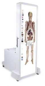
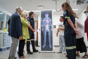
The newest version displays interactive, 3-D imagery of human anatomies on a life-size screen. Anatomage image.
The table is the latest release from a U.S.-based developer of medical imaging technology called Anatomage. And there are several now in operation here in Canada.
The newest version, Table 10, displays interactive, 3-D imagery of human anatomies on a life-size screen (measuring 2.13 by 0.67 metres, screens can be used in vertical or horizontal mounts). And the ‘bed’ on which the ‘patient’ is lying is like a giant smartphone! It’s interactive, interconnected, touch-sensitive and can be upgraded through software to deliver authentic, high-definition visualizations of various human bodies with differing medical conditions.
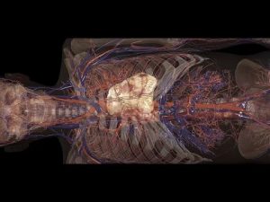
Anatomage devices have the giant touchscreen so learners can ‘pull things apart’ virtually to expand, shift or rotate anatomical features displayed on-screen. Body tissues or systems can be added or removed as needed; muscles, bones, connective tissues, arteries and vessels can be magnified, manipulated or even surgically dissected and segmented virtually by a user.
There’s a comprehensive real anatomy database of imagery assembled for education and training purposes, and various elements of the digital cadavers displayed on the table can be digitally enhanced to magnify important details.
For example, the human cardiovascular system, including arteries and veins, is fully traced and functionally connected in this virtual world. Each structure is rendered in ultra-high resolution, down to 200 microns, providing an unprecedented level of anatomical detail.
Familiar medical imaging technologies (like CT scan, X-ray, ultrasound, and MRI) are integrated into the table’s touchscreen interface, while advanced features like endoscopic fly-throughs and ultrasound viewers are also incorporated to simulate real-world clinical experiences.
Embedded software existing in the tables comes with four cadavers – two male and two female. They are Caucasian and Asian, and their case histories come complete with gross anatomy images, case studies, and several pathology and histology slides (tens of thousands are available). The images are from non-chemically treated cadavers, with colour and shape well-preserved.
California-based Anatomage worked with the Clinical Anatomy Department at Stanford University to develop the device for instructional and diagnostic purposes, and imagery featured on the platform originated and was licensed from cadavers included in what’s known as the Visible Korean Human (VKH) and Visible Human Project (VHP).
The company has in its wide range of medical assets and reconstructed human anatomies a fully interactive 3-D simulation of the childbirth process, too. There’s also a dental visualization product, and a veterinarian table for study and treatment of animals.
Cadaver imagery featured on the platform originated and was licensed for instructional and diagnostic purposes.
The latest Table 10 software introduces Anatomage’s newest cadaver, called Hans, reconstructed from an actual 70-year-old patient who passed away from lung cancer. Hans showcases an intricate muscular and vascular system, giving practitioners a detailed look at geriatric anatomy.
Students can examine Hans to discover the natural aging process, and to gain visual insights into conditions commonly affecting older populations, including metastasized cancer and tumours in the liver, pancreas and chest wall, promoting understanding and treatment of patients in this age group.
At the other end of the life cycle, a Birth Simulation program is another addition to Table 10, providing a fully interactive, anatomically-correct simulated birth. Visualizing childbirth and delivery is challenging without detailed 3-D views from multiple angles that show anatomical transformation taking place during the different stages of labour, including cervical dilation, infant rotation and head movements, and the release of the placenta.
And with the latest acquisition of Anatomage high-tech tables by top Canadian teaching institutions, more and more medical students, educators, and professionals here can interact with, and learn from, not just a cadaver of an older adult impacted by cancer or complications encountered in human childbirth.
Students at Conestoga College are now working with an Anatomage table. Image from Conestoga College.
Students at Conestoga College, who are training to be a paramedic, nurse, personal support worker, medical lab technician, pharmacy technician, recreational therapist, occupational therapist, massage therapist or other health care professional, can now work with an Anatomage table – four of them in fact, along with other updated facilities, curated labs and supportive technologies that have been installed to meet student education needs.
“These tables will provide Conestoga the opportunity to enhance learning and help bridge knowledge gaps across all the various health sciences programs within lab and during independent practice,” Kyla Rotobilsky, manager of simulation and learning innovation, said when the acquisition was announced.
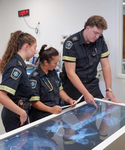
A simulated scenario of a double gunshot victim was part of the the Anatomage Table demonstration at Conestoga College.
Describing a recent demonstration, Rotobilsky explained how an Anatomage table was integrated into a simulated scenario of a double gunshot victim alongside a high-fidelity mannequin. Paramedic students assessed the simulated patient and identified critical steps in the care needed.
Once the patient was stabilized, the students moved to the Anatomage table where they could further analyze the impact of the trauma. The integrated scenarios built within the table allowed the students to examine and highlight a detailed X-ray image to determine potential further damage to different systems of the body caused by the gunshot wounds.
Conestoga’s acquisition brings the sophisticated medical training technology to a handful of Canadian universities, including St Mary’s, Memorial, Brock, Simon Fraser, and others.
Students at these and other such institutions have a fun way to continue their learning by joining in an Anatomage Tournament, an exciting mash-up of medical education and 3-D gamification.
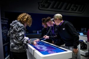
Every year, hundreds of students gather round tables in local tournaments, leading up to the National Anatomage Tournament.
The Anatomage Tournament tests students’ understanding of anatomical structures using the Anatomage table as they interact with a digitized human cadaver—rotating, zooming in, and tapping on structures to select correct answers on anatomy and physiology.
Every year, hundreds of students gather ’round tables in local tournaments, leading up to the National Anatomage Tournament.
At the top of their ‘game’ during a recent Tournament, Canadian medical students were on the winning team, taking first place in a competition that can have real world life-and-death consequences!
-30-